

 journal menu


issue contents
October 2024 issue
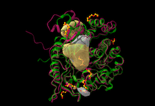
Cover illustration: β-Glucosidase from the thermophilic bacterium Caldicellulosiruptor saccharolyticus (Bgl1) [Sotiropoulou et al. (2024), Acta Cryst. D80, 733–743]. Superposition of the crystal structures of Bgl1 (red) and β-glucosidase from C. cellulovorans (green). The catalytic site of Bgl1 and an additional adjacent cavity that was detected by the DeepSite algorithm are highlighted in surface representation (yellow).
CCP4


Open  access
access
This article examines the application of anomalous scattering for the identification of elements within crystal structures of macromolecules.
research papers

Open access
access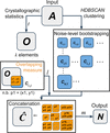
Beamstops and beamstop shadows can lead to erroneous reflection measurements in crystallographic diffraction experiments, which is a common problem in data processing. Here, a method is presented that combines crystallographic statistics with machine learning to robustly detect such reflections.

Open access
access
Crystal structures of a β-glucosidase from the thermophilic bacterium C. saccharolyticus (Bgl1) and its complex with glucose were determined at 1.47 and 1.95 Å resolution, respectively. Comparison of Bgl1 with sequence or structural homologues showed that Bgl1 is quite similar except for two regions, one of which seems to be a unique insertion of residues that is only present in Bgl1.
Open access
access
A description is given of new deep-learning tools that analyse experimental micrographs of crystallization experiments to enable the automation of outcome classification, crystal detection and determination of locations to dispense compounds for fragment-based drug discovery at Diamond Light Source.


