journal menu


issue contents
November 2020 issue
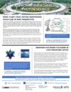
Cover illustration: Reconstruction of human pancreatic tissue, showing the islet of Langerhans, a cluster of cells important in endocrine (hormone-producing) function: (upper left) cut through the reconstruction volume, (upper right) segmentation into different tissue components,(lower left) spatial arrangments of the cluster, (lower right) validation of segmentation and selection of cells for further analysis; scalebar: 50 µm. The sample was collected during a pancreatic tumor surgery. The paper by Frohn, Pinkert-Leetsch, Missbach-Güntner, Reichardt, Osterhoff, Alves and Salditt (pages 1707-1719) demonstrates how phase contrast tomography combining parallel and cone beam recording geometries can help to perform quantitative 3D analysis for virtual histopathology.
facility information

international union of crystallography


 access
 access
research papers
 access
 access
 access
 access

Geometric determination of direction of dislocations using synchrotron X-ray transmission topography
access access
Zernike phase-contrast full-field transmission X-ray nanotomography for 400 micrometre-sized samples
access access
short communications
 access
computer programs
access
obituaries